The Cardiovascular System
The Blood
The Heart |
Cardiac Cycle |
The Blood |
Types of Arteries |
Blood Circulation |
Blood Pressure |
ECG

In the case of the left ventricle, the afterload is a consequence of the blood pressure, since the pressure in the ventricle must be greater than the blood pressure in order to open the aortic valve. The fluid pumped by the heart that circulates throughout the body via the arteries, veins, and capillaries. An adult male of average size normally has about 6 quarts (5.6 liters) of blood. The blood carries oxygen and nutrients to the body tissues and removes carbon dioxide and other wastes. The colorless fluid of the blood, or plasma, carries the red and white blood cells, platelets, waste products, and various other cells and substances.
The blood Vessels
The blood vessels are elastic tubular canals through which blood circulates in the body. Different types of blood vessels include Arteries, veins and Capillaries.
The blood vessels are part of the circulatory system and function to transport blood throughout the body. The most important types, arteries and veins, are so termed because they carry blood away from or towards the heart, respectively. The arteries carry blood away from the heart; the main arterial vessel, the aorta, branches into smaller arteries, which in turn branch repeatedly into still smaller vessels and reach all parts of the body. Within the body tissues, the vessels are microscopic capillaries through which gas and nutrient exchange occurs. Blood leaving the tissue capillaries enters converging vessels, the veins, to return to the heart and lungs.
Structure of Arterial Wall
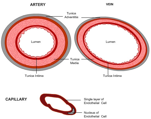
All blood vessels follow the same histological makeup. The arterial layer that is in direct contact with the flow of blood is the tunica intima, commonly called the intima. This layer is made up of mainly endothelial cells. Just deep to this layer is the tunica media, known as the media. This "middle layer" is made up of smooth muscle cells and elastic tissue. The outermost layer (furthest from the flow of blood) is known as the tunica adventitia or the adventitia. This layer is composed of connective tissue.

Tunica intima or tunica interna
The tunica intima is the inner layer of arteries and veins. In arteries this layer is composed of an elastic membrane lining and smooth endothelium that is covered by elastic tissues. A layer of endothelial cells covers the lumen of the vessel, as well as a sub-endothelial layer made up of mostly loose connective tissue. Often, the internal elastic lamina separates the tunica intima from the tunica media.
This layer is comprised of a smooth lining of endothelial cells, which also forms the valves, and a basement membrane. Due to this layer the arteries are able to cope with the high-pressure surges of blood from the heart because they have very thick muscular walls. The single layer of cells that make up a capillary enables the passage of oxygen and nutrients from capillaries into the surrounding tissue. Veins have loose, slack walls as the blood inside them is under very little pressure.
The blood vessels are capable of either constricting (Vasoconstriction) and decreasing the lumen size or dilating (vasodilatation) and increasing the lumen size. Vasoconstriction is the major contributing factor leading to high blood pressure.
Capillaries consist of little more than a layer of endothelium and occasional connective tissue.
Types
Blood vessels exist in varying calibers:
- Arteries
- Aorta (the largest artery, carries blood out of the heart)
- Branches of the aorta, such as the carotid artery, the subclavian artery, the celiac trunk, the mesenteric arteries, the renal artery and the ileac artery.
- Arterioles
- Capillaries (the smallest blood vessels)
- Venules
- Veins
- Large collecting vessels, such as the subclavian vein, the jugular vein, the renal vein and the iliac vein.
- Venae cavae (the 2 largest veins, carry blood into the heart)
They are roughly grouped as arterial and venous, determined by whether the blood in it is flowing toward or away from the heart. The term "arterial blood" is nevertheless used to indicate blood high in oxygen, although the pulmonary artery carries "venous blood" and blood flowing in the pulmonary vein is rich in oxygen.

 Types of Arteries